- Shortness of breath since 5 days
- Chest pain since 5 days
- With no other significant complaints
the history reveled the patient had similar complaints 3 times in last two years and each time he was treated with tube thoracostomy. only 30 days back he was treated with tube thoracostomy which was removed 7 days later and patient was fine
 |
| 30 days back |
 |
| after tube thoracostomy |
Now he has again developed same complaints. Patient was a mild smoker for 5 years, smoked only 3-5 cigarettes/ day and stopped 3 years back when he had first time pneumothorax.
ON GENERAL EXAMINATION:
General physical examination was all normal except for mild dyspnea.
RESPIRATORY EXAMINATION:
- Shape of chest- Normal
- Type of respiration – Abdominothoracic
- Trail’s sign – Positive - towards right side
- Apex beat – Shifted towards mid line
- 3 Scars were present in mid axillary line on left side
- Chest expansion – Reduced on left side
- Chest movement - Reduced on left side
- A/E absent on left side in mid axillary
- VR and VF both decreased
- Hyper-resonant note on left side of lung
CARDIOVASCULAR SYSTEM:
S1 and S2 heard
no murmur
CENTRAL NERVOUS SYSTEM:
NAD
P/A:
soft
no organomegaly
CHEST RADIOGRAGH:
TREATMENT: treatment with tube thracostomy was done with good antibiotic coverage, incentive spirometery and symptomatic treatment was also started and BPF (bronchopleural fistula) was also present
PLEUROSCOPY:
It showed multiple adhesions between viceral and perital pleura with few sub pleural blebs.
but after tube thoracostomy patient was quite well and he started to move around with tube in situ , at this point of time he was discharged.
Then after a weeks time he turned back and this time with tube in situ, column moving and
cheif complaint of
 |
| on admission |
TREATMENT: treatment with tube thracostomy was done with good antibiotic coverage, incentive spirometery and symptomatic treatment was also started and BPF (bronchopleural fistula) was also present
PLEUROSCOPY:
It showed multiple adhesions between viceral and perital pleura with few sub pleural blebs.
but after tube thoracostomy patient was quite well and he started to move around with tube in situ , at this point of time he was discharged.
Then after a weeks time he turned back and this time with tube in situ, column moving and
cheif complaint of
- high grade fever
- dyspnea since 1 day
• BP – 120/70 mm hg
• Pulse – 102/ min
• RR – 28/ min
RESPIRATORY EXAMINATION:
• Left sided ICD functional, column moving
• Shape of chest – right side chest hyper inflated
• Trail’s sign – positive
• Trachea shifted to left side
• A/E present on left side
• A/E absent on right side
• Hyper resonant note on right side
Immediately on the basis of clinical examination we suspected right sided pneumothorax and
2 wide bore needles were inserted in the intercostal space and patient was urgently sent for X-ray chest. |
| left side ICD and right side pneumothorax |
This time patient was having pneumothrax on right side and we had to do tube thoracostomy on right side.
now for me this was for the first time when i had done tube thoracostomy on the both side of the patient and now patient was suffering from BILATERAL PNEUMOTHORACES with ICDs inserted to the both sides. patients alpha 1 anti tripsin levels were adviced and it came out to be on higher side( 600) ruling out the posibility of alpha 1 antitripsin defficiency, patient was in vestigated for other cause of reccurent pneumothoraces but every investigation was within normal range, patient was HIV negative.
 |
| ICD in both pleural spaces |
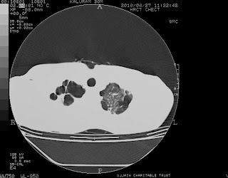
Patient started improving slowly and we advised the patient for CT Thorax, it was advised in very beginning but patient was not able to afford the expenses. He now agreed to pay for his CT and the following reading was noted on CT scan of
 |
| large bullae in both apice of lung |
 |
| multiple bullae |
 |
| multiple bullae |
 |
| bullae in both lungs |
 |
| multiple bullae in upper lobes of both lungs |
The CT of the patient was having multiple emphysematous bullae in upper and middle lobe of right lung with upper lobe involvement in left lung. The bullae was more on right side but the patient had 7 times pneumothorax on left side. Diagnosis of formation of bullae in lungs due to smoking, but formation of bullae due to such a short period of exposure to smoking and to that much less amount of cigarettee smoking was very unconvincing or rather we shall say was VERY RARE.

